|
Protozoan and Helminth Parasites
Contents
 Macrophages are one of the main phagocytic cells capable of destroying parasites. Once the parasite is ingested by these cells, they begin to produce nitric oxide (NO), a potent chemical that is toxic to these pathogens. In fact, NO related products are toxic to malaria, leishmania, trypanosomes, shistosome larvae. interferon-gamma (IFN-γ) activates the macrophage that produces the NO (Figure 3). Macrophages are one of the main phagocytic cells capable of destroying parasites. Once the parasite is ingested by these cells, they begin to produce nitric oxide (NO), a potent chemical that is toxic to these pathogens. In fact, NO related products are toxic to malaria, leishmania, trypanosomes, shistosome larvae. interferon-gamma (IFN-γ) activates the macrophage that produces the NO (Figure 3).
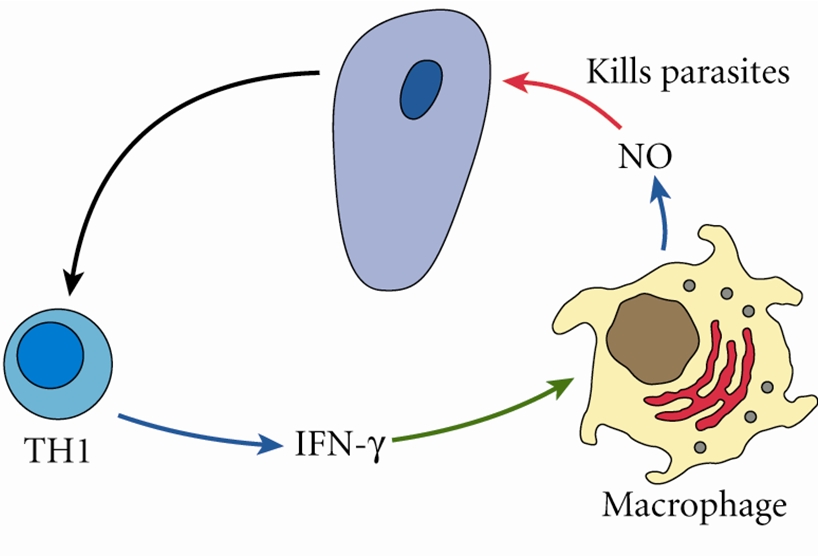
Figure 3. Direct killing by cytokine-activated macrophages. TH1 (T Helper 1) cells generates a signal (IFN-γ). IFN-γ activates the macrophage. The macrophage can either secrete NO to destroy the pathogen or release the NO within its cytoplasm to directly destroy ingested pathogens (not shown). The arrow pointing from the parasite to the TH1 cell indicates that it is the presence of the parasite within the host that activates the TH1 cell. Click to enlarrge.
 Antigens of many intracellular parasites are processed and presented via the endogenous antigen-presenting pathway. This involves ingesting the parasite, digestion within the phagocyte's cytoplasm, and presenting using MHC class I molecules. Antigens presented by MHC class I molecules become targets for cytotoxic T lymphocytes (CTL). CTL like their targets by inserting perforin protein into the cell membrane of the infected cell presenting the parasitic antigen, essentially creating a pore-like hole in the membrane. The CTL then releasing granzymes (specialized digestive enzymes) into the infected cell, which induces apoptosis to occur shortly after. The CTL detaches from its target and goes to look for another infected cell (Figure 4). Antigens of many intracellular parasites are processed and presented via the endogenous antigen-presenting pathway. This involves ingesting the parasite, digestion within the phagocyte's cytoplasm, and presenting using MHC class I molecules. Antigens presented by MHC class I molecules become targets for cytotoxic T lymphocytes (CTL). CTL like their targets by inserting perforin protein into the cell membrane of the infected cell presenting the parasitic antigen, essentially creating a pore-like hole in the membrane. The CTL then releasing granzymes (specialized digestive enzymes) into the infected cell, which induces apoptosis to occur shortly after. The CTL detaches from its target and goes to look for another infected cell (Figure 4).
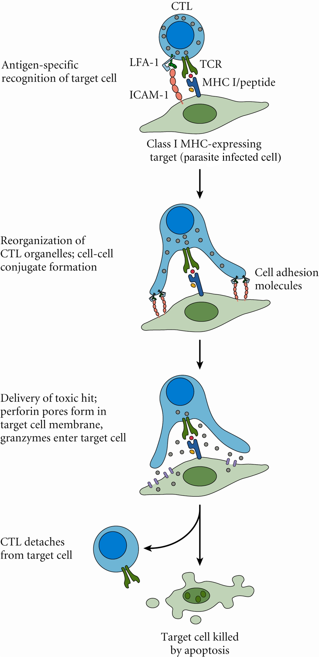
Figure 4. Stages of cytotoxic T lymphocyte cytotoxicity.
 Antibodies: Important for some protozoa's and helminths that infect the gut (Figure 5). Antibodies: Important for some protozoa's and helminths that infect the gut (Figure 5).
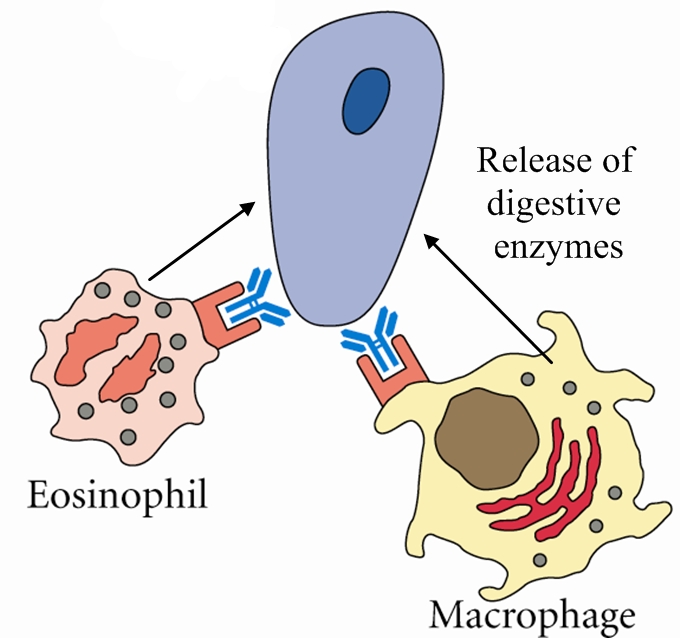
Figure 5. Antibodies produced by the humoral part of the immune system (B lymphocytes) bind to parasites found in the gut. The Fc portion of the antibody then bind to FcR (Fc receptor) on macrophages and eosinophils which activate them to produce and release toxic chemicals that kill the parasite. This is an example of antibody-dependent cellular cytotoxicity (ADCC).
Click to enlarge.
 Mast cells and goblet cells for intestinal parasites. Mast cells release toxic mediators, while mucin secretions by goblet cells prevents nutrient uptake by the parasite and may aid in flushing out the parasite. IL-4 may also induce gut motility. Mast cells and goblet cells for intestinal parasites. Mast cells release toxic mediators, while mucin secretions by goblet cells prevents nutrient uptake by the parasite and may aid in flushing out the parasite. IL-4 may also induce gut motility.
 Eosinophils are important for destroying larval stages of helminth parasites. Eosinophils are important for destroying larval stages of helminth parasites.


The following is a list of parasites that have been profiled:
|
