Vibrio parahemolyticusVibrio parahaemolyticus is a Gram-negative, rod-shaped, non-spore-forming, highly motile bacterium possessing all the typical components of Gram-negative bacteria, such as peptidoglycan, O-antigen repeating subunits, core polysaccharide, lipopolysaccharide, and lipid A anchor (Figure 1). It is a naturally occurring pathogen widely disseminated in marine and estuarine environments of tropical and temperate areas that causes mild or moderate gastroenteritis with symptoms of diarrhea, abdominal cramps, nausea, vomiting, headache, fever and chills. V. parahaemolyticus grow optimally under high salt concentrations, and is therefore referred to as a halophilic species (Daniels et al., 2000). However, during the warmer seasons, an increase in seawater temperature causes the number of V. parahaemolyticus bacteria in coastal regions to increase as well, making humans highly susceptible to exposure and infection. V. parahaemolyticus is known to get into the human body mainly through the consumption of undercooked or raw seafood, such as oysters or shellfish, but exposure to wounded flesh can also provide the pathogen access to targeted organs (Ryan & Ray, 2004). It is estimated that one in every 400000 individuals are affected by the bacteria; thus, while the risks are relatively low, it is important to always have your seafood cooked thoroughly when dining out.
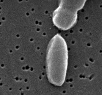
Figure 1. This scanning electron micrograph depicts a Vibrio parahaemolyticus bacterium [19058 X]. V. parahaemolyticus exhibits high motility due to its flagella, which are located at the polar ends of the bacterium cell. The flagellum functions to propel the organism through liquid medium. A single flagellum is typically more effective in liquids that are less dense; the opposite is true for polar flagella, which are generally more effective in denser, more viscous liquids (McCarter, 1999). Some strains of V. parahaemolyticus, however, possess a single flagellum (Figure 1). Furthermore, V. parahaemolyticus uses oxygen as its final electron acceptor when generating ATP via cellular aerobic respiration, and is therefore classified as oxidase-positive. Although, the major source of energy production for V. parahaemolyticus is through the electron transport chain, it can also produce ATP in the absence of oxygen; hence, this species is a facultative anaerobe.
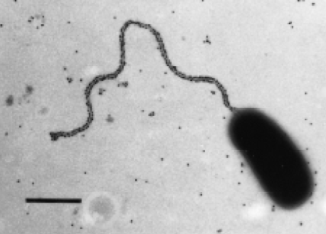
Figure 2. Electron micrograph of a V. parahaemolyticus bacterium [Bar = 1 μm]. The presence of V. parahaemolyticus can be diagnosed by isolating the bacteria from a sample of infected stool or blood. Compounds such as blood agar or thiosulfate can be used to culture the pathogen (Figure 3); the latter compound is often recommended under clinical setup.
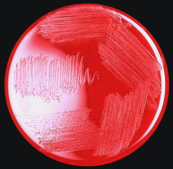
Figure 3. Blood agar plate culture of Vibrio parahaemolyticus. Watery diarrhea, vomiting, abdominal cramps, headache, nausea, and other signs related to gastrointestinal diseases are the normal symptoms found in a person infected with V. parahaemolyticus (Daniels et al. 2000). The major key virulence factors associated with V. parahaemolyticus is TDH (thermostable direct hemolysin) and TRH (thermostable direct hemolysin-related hemolysin). TDH is known to exhibit the Kanagawa phenomenon, which is involved in the β-hemolysis of the red blood cells present in humans (McLaughlin et al., 2005). No long-term illnesses are related to V. parahaemolyticus infection, although a few deaths have been documented in places such as Australia. The extent of illness or infection induced by V. parahaemolyticus depends on the amount of infected food ingested. References: Daniels, N.A., Ray, B., et al. (2000). Emergence of a new Vibrio parahaemolyticus serotype in raw oysters: A prevention quandary. Journal of the American Medical Association, 284(12): 1541-1545. McCarter, L. (1999). The Multiple Identities of Vibrio parahaemolyticus. Journal of Molecular Microbiology and Biotechnology, 1(1): 51-57. McLaughlin, J.B., DePaola, A., et al. (2005). Outbreak of Vibrio parahaemolyticus gastroenteritis associated with Alaskan oysters. The New England Journal of Medicine, 353(14): 1463-1470. Ryan, K.J.; Ray, C.G. (2004). Sherris Medical Microbiology (4th ed.). New York: McGraw Hill. |