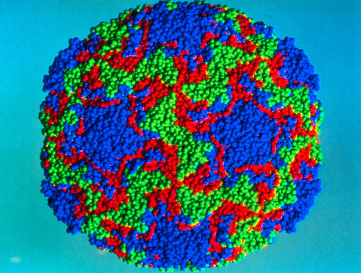
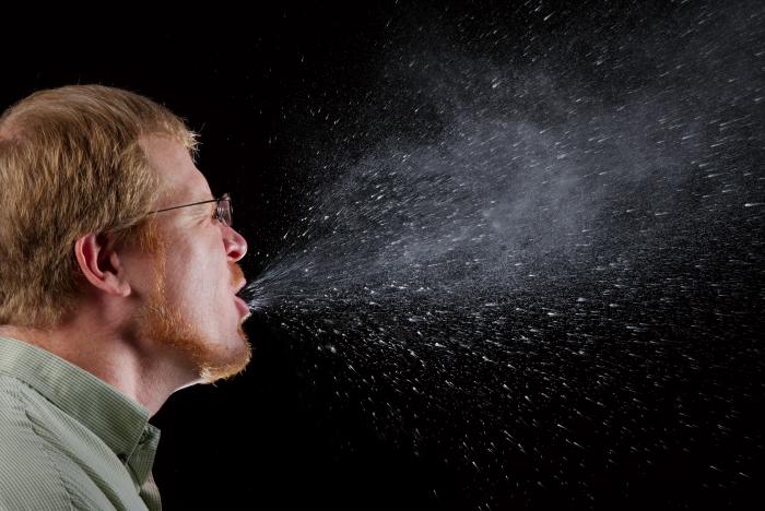
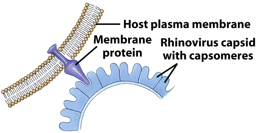
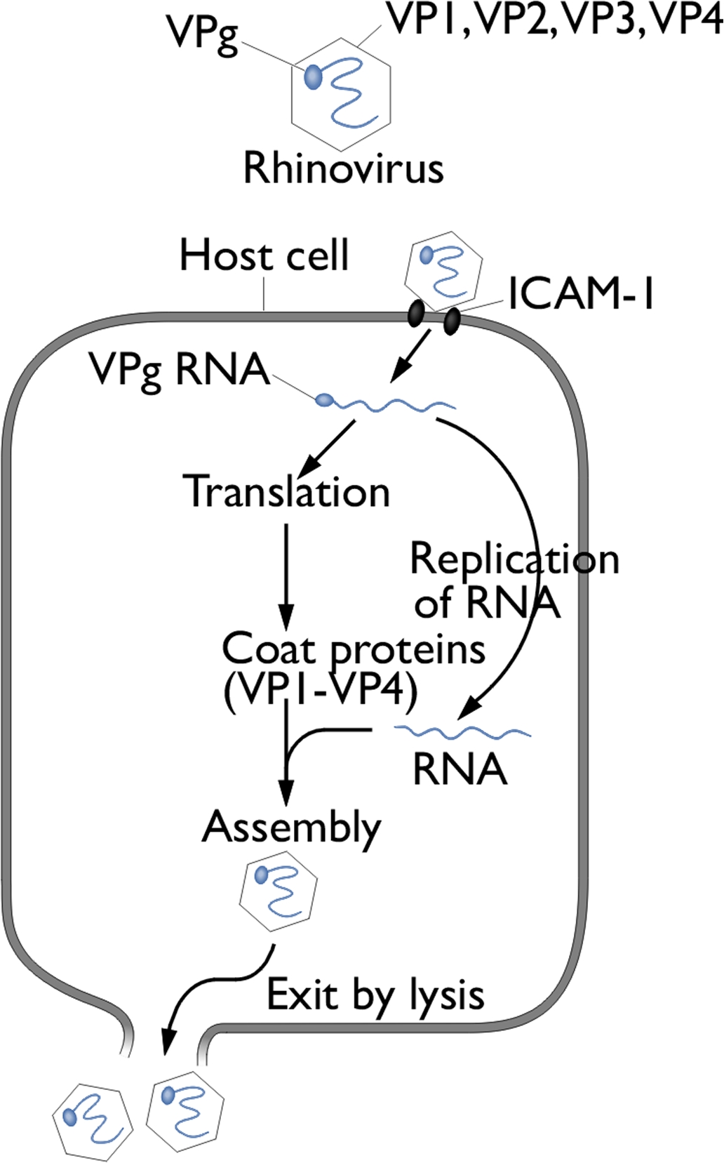
RhinovirusOverview: Rhinovirus is a single-stranded, positive-sense RNA virus that possesses a naked (as opposed to enveloped) icosahedral assembly of 60 protomers (Figure 1). There are 100 recognized serotypes of this particular virus, making it the most common viral agent in humans, and a causative agent of the common cold. Due to its small size and diameter of about 30 nm, this pathogen's genome is approximately 7.2 to 8.2 kilobases in length and possesses a virus encoded protein at its 5’ (five prime) end and a poly(A)-tail at its 3' end. The capsid is composed of four viral proteins: VP1, VP2, VP3, and VP4. The viral proteins VP1, VP2, and VP3 are epitopes that interact with antibodies of the host's immune system (Larralde et al., 1991). Figure 1. A graphical representation of rhinovirus. Pathogenicity: Rhinovirus can be transmitted by two methods, namely, via respiratory droplets and from contaminated surfaces, including direct person-to-person contact (Figure 2). The primary method of entrance into the body is through the nasal mucosa. Optimal replication of rhinovirus occurs between 33°C to 35°C; this is why viral replication occurs best in the nasal passages and upper tracheobronchial trees, as opposed to the lower respiratory tract, which is close to 37°C. Figure 2. This photograph demonstrates a sneeze in progress, revealing the plume of salivary droplets as they are expelled in a large cone-shaped array from this man’s open mouth, thereby, dramatically illustrating the reason one needs to cover his/her mouth when coughing, or sneezing, in order to protect others from germ exposure. When a rhinovirus virion first encounters targeted epithelial cells of the upper respiratory tract, it attaches to intracellular adhesion molecule-1 (ICAM-1) on respiratory epithelium via VP1 and enters by endocytosis (Figure 3). Viral RNA is then synthesized and translated into VP1 through VP4. Since the virus initially contains a positive-sense RNA strand as its genome, the RNA is used as a template to produce a negative-sense RNA strand. This is done using viral replicase, an enzyme which is usually carried in the nucleoprotein core. The negative-sense strand is subsequently used as a template to generate many strands of positive-sense RNA, as the genome for progeny virions being produced (Figure 4). The assembly of viral proteins and RNA creates new virions that exit the infected cell by lysis and spread locally to invade neighboring cells. The replication cycle of rhinovirus is summarized in Figure 5.
Figure 3. The attachment of rhinovirus capsid proteins to target cell membrane proteins. The crevice in which the membrane protein fits into is called the 'canyon'.
Figure 4. Overview of viral replication strategies. Click to enlarge. Figure 5. Structure & replication cycle of rhinovirus. Symptoms of Infection: Incubation periods last about two to four days. Inflammatory responses caused by rhinovirus in the respiratory tract can lead to:
At the moment, there is no true vaccine or treatment available to prevent or treat infections associated with the virus, other than a few antiviral drugs that inhibit viral RNA from being injected into the host, such as pleconaril (Heikkinen et al., 2003) . Research conducted by Larralde et al. (1991) showed that even though there are many serotypes of rhinovirus, similarities in the VP4 protein give hope for future vaccines. References: Keikkinen, T., & Jarvinen, A. (2003). The common cold. Lancet, 361: 51-59. Larralde, G., Li, B., Kapikian, A., & Gorziglia, M. (1991). Serotype-specific epitope(s) present on the vp8 subunits of rotavirus vp4 protein. Journal of Virology, 65(6): 3213-3218. |